Research News
Jun 3, 2026
- Veterinary Science
Computed tomography findings of pulmonary lymphoma in a dog and two cats
CT of pulmonary lymphoma
Distinct pulmonary vessels, indicated with triangles, and air bronchograms, represented with arrows, were observed in lesions.
Credit: Osaka Metropolitan University
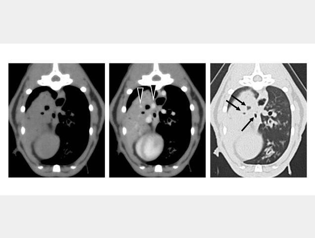
In a case series, an Osaka Metropolitan University-led research team assessed CT findings of pulmonary lymphoma in a dog and two cats. The dog (Case 1) showed defined consolidation and nodules with enlarged sternal and mediastinal lymph nodes. Air bronchogram and distinct pulmonary vessels were observed within the lesion. Several nodules were also found in the kidneys. Pulmonary and kidney lesions were diagnosed as B cell lymphoma.
One cat (Case 2) showed well-defined consolidation with an enlarged tracheobronchial lymph node. Air bronchogram and distinct pulmonary vessels were also observed in the lesion. No additional lesions were detected. The pulmonary lesion was diagnosed as B cell lymphoma. Another cat (Case 3) showed a nasopharyngeal mass and multiple well-defined pulmonary nodules without lymph adenomegaly. Pulmonary nodules and the nasopharyngeal mass were diagnosed as lymphoma. In Case 3, T/B classification was not performed.
Of three pulmonary lymphoma cases, the distinguishing imaging feature was a well-defined lesion with distinct air bronchograms, pulmonary vessels, and homogeneous enhancement.
Paper information
Journal: Veterinary Medicine and Science
Title: Computed Tomography Findings of Pulmonary Lymphoma in a Dog and Two Cats
DOI: 10.1002/vms3.70947
Authors: Toshiyuki Tanaka, Yusuke Wada, Takuya Kusaka, Mizuki Tomihari, Takashi Hasegawa
Published: 7 April 2026
URL: https://doi.org/10.1002/vms3.70947
Contact
Toshiyuki Tanaka
Graduate School of Veterinary Science
Email: t-tanaka[at]omu.ac.jp
*Please change [at] to @.
SDGs
