Research News
Jul 7, 2023
- Medicine
AI finds a way to people’s hearts (literally!)
Unveiling a groundbreaking and accurate AI-based method to classify cardiac function and disease using chest X-Rays
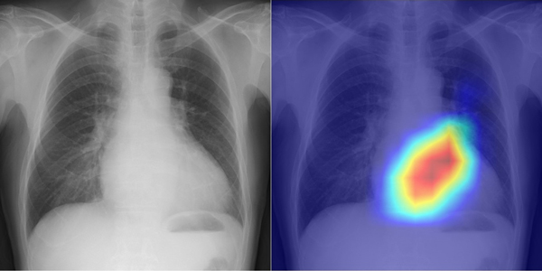
Evaluation of Left Ventricular Ejection Fraction
Left: Chest radiograph
Right: Visualization of the grounds for the AI's judgment
Credit: Daiju Ueda, OMU
Osaka, Japan - AI (artificial intelligence) may sound like a cold robotic system, but Osaka Metropolitan University scientists have shown that it can deliver heartwarming—or, more to the point, “heart-warning”—support. They unveiled an innovative use of AI that classifies cardiac functions and pinpoints valvular heart disease with unprecedented accuracy, demonstrating continued progress in merging the fields of medicine and technology to advance patient care. The results will be published in The Lancet Digital Health.
Valvular heart disease, one cause of heart failure, is often diagnosed using echocardiography. This technique, however, requires specialized skills, so there is a corresponding shortage of qualified technicians. Meanwhile, chest radiography is one of the most common tests to identify diseases, primarily of the lungs. Even though the heart is also visible in chest radiographs, little was known heretofore about the ability of chest radiographs to detect cardiac function or disease. Chest radiographs, or chest X-Rays, are performed in many hospitals and very little time is required to conduct them, making them highly accessible and reproducible. Accordingly, the research team led by Dr. Daiju Ueda, from the Department of Diagnostic and Interventional Radiology at the Graduate School of Medicine of Osaka Metropolitan University, reckoned that if cardiac function and disease could be determined from chest radiographs, this test could serve as a supplement to echocardiography.
Dr. Ueda’s team successfully developed a model that utilizes AI to accurately classify cardiac functions and valvular heart diseases from chest radiographs. Since AI trained on a single dataset faces potential bias, leading to low accuracy, the team aimed for multi-institutional data. Accordingly, a total of 22,551 chest radiographs associated with 22,551 echocardiograms were collected from 16,946 patients at four facilities between 2013 and 2021. With the chest radiographs set as input data and the echocardiograms set as output data, the AI model was trained to learn features connecting both datasets.
The AI model was able to categorize precisely six selected types of valvular heart disease, with the Area Under the Curve, or AUC, ranging from 0.83 to 0.92. (AUC is a rating index that indicates the capability of an AI model and uses a value range from 0 to 1, with the closer to 1, the better.) The AUC was 0.92 at a 40% cut-off for detecting left ventricular ejection fraction—an important measure for monitoring cardiac function.
“It took us a very long time to get to these results, but I believe this is significant research,” stated Dr. Ueda. “In addition to improving the efficiency of doctors’ diagnoses, the system might also be used in areas where there are no specialists, in night-time emergencies, and for patients who have difficulty undergoing echocardiography.”
Funding
None.
Paper Information
Journal: The Lancet Digital Health
Title: Artificial intelligence-based model to classify cardiac functions from chest radiographs: a multi-institutional, retrospective model development and validation study
DOI: 10.1016/S2589-7500(23)00107-3
Author: Daiju Ueda, Toshimasa Matsumoto, Shoichi Ehara, Akira Yamamoto, Shannon Leigh Walston, Asahiro Ito, Taro Shimono, Masatsugu Shiba, Tohru Takeshita, Daiju Fukuda, Yukio Miki
Publication date: July 6, 2023 (BST)
https://www.thelancet.com/journals/landig/article/PIIS2589-7500(23)00107-3/fulltext
Contact
Graduate School of Medicine
Daiju UEDA
E-mail: ai.labo.ocu[at]gmail.com
*Please change [at] to @.
SDGs
