Research News
Oct 6, 2023
- Science
Super-efficient laser light-induced detection of cancer cell-derived nanoparticles
Skipping ultracentrifugation, detection time reduced from hours to minutes!
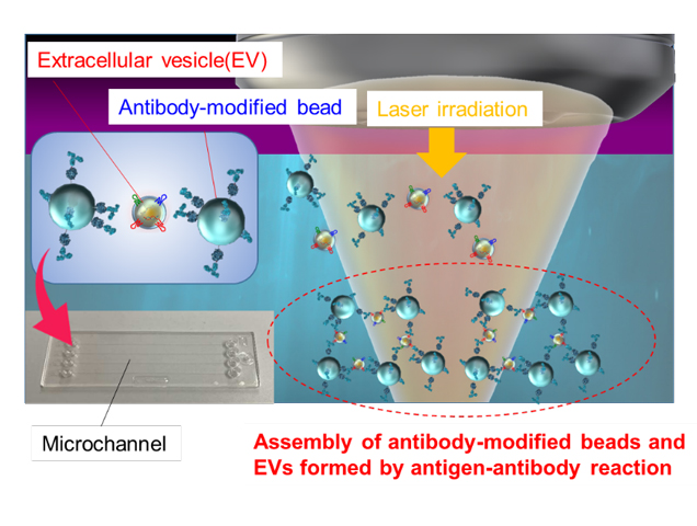
Schematic diagram of light-induced assembly of extracellular vesicles (EV)
Using laser irradiation, the researchers managed to directly detect nanoscale EVs in a cell supernatant within minutes.
Credit: Takuya Iida, Osaka Metropolitan University
Osaka, Japan - Can particles as minuscule as viruses be detected accurately within a mere 5 minutes? Osaka Metropolitan University scientists say yes, with their innovative method for ultrafast and ultrasensitive quantitative measurement of biological nanoparticles, opening doors for early diagnosis of a broad range of diseases.
Nanoscale extracellular vesicles (EVs) including exosomes, with diameters of 50–150 nm, play essential roles in intercellular communication and have garnered attention as biomarkers for various diseases and drug delivery capsules. Consequently, the rapid and sensitive detection of nanoscale EVs from trace samples is of vital importance for early diagnosis of intractable diseases such as cancer and Alzheimer's disease. However, the extraction of nanoscale EVs from cell culture media previously required a complex and time-consuming process involving ultracentrifugation.
A research team led by Director Professor Takuya Iida, Deputy Director Associate Professor Shiho Tokonami, and Assistant Director Professor Ikuhiko Nakase, from the Research Institute for Light-induced Acceleration System (RILACS) at Osaka Metropolitan University, has utilized the power of laser light to accelerate the reaction between nanoscale EVs derived from cancer cells and antibody-modified microparticles. The three-dimensional structure of the resulting aggregates was then analyzed using confocal microscopy. As a result, the researchers demonstrated the ability to measure, within 5 minutes, approximately 103–104 nanoscale EVs contained in a 500 nL sample.
Professor Iida concluded, “This research achievement provides a method for ultrafast and ultrasensitive quantitative measurement of biological nanoparticles, offering a foundation for innovative analysis of cell-to-cell communication and early diagnosis of various diseases in the future.”
Their findings were published in Nanoscale Horizons.
Funding
This study was supported by the JST-Mirai Program (No. JPMJMI18GA, No. JPMJMI21G1), Grant-in-Aid for Scientific Research (A) (No. JP17H00856, No. JP21H04964), JST FOREST Program (No. JPMJFR201O), Grant-in-Aid for Scientific Research (B) (No. JP18H03522), Scientific Research on Innovative Areas (No. JP16H06507), Grant-in-Aid for Early-Career Scientists (No. JP20K15196) from Japan Society for the Promotion of Science KAKENHI, and the Key Project Grant Program of Osaka Prefecture University.
Paper Information
Journal: Nanoscale Horizons
Title: Ultrafast sensitivity-controlled and specific detection of extracellular vesicles using optical force with antibody-modified microparticles in a microflow system
DOI: 10.1039/d2nh00576j
Author: Kana Fujiwara, Yumiko Takagi, Mamoru Tamura, Mika Omura, Kenta Morimoto, Ikuhiko Nakase, Shiho Tokonami and Takuya Iida
Published: 12 Jul 2023
URL: https://doi.org/10.1039/d2nh00576j
Contact
Graduate School of Science
Takuya IIDA
E-mail t-iida[at]omu.ac.jp
Shiho TOKONAMI
E-mail tokonami [at]omu.ac.jp
Ikuhiko NAKASE
E-mail i-nakase[at]omu.ac.jp
*Please change [at] to @.
SDGs

