Research News
Aug 26, 2025
- Veterinary Science
Making low-fertility rats fertile by changing the treatment interval
Changing the interval between fertility drugs boosts fertility even in rats that typically respond poorly
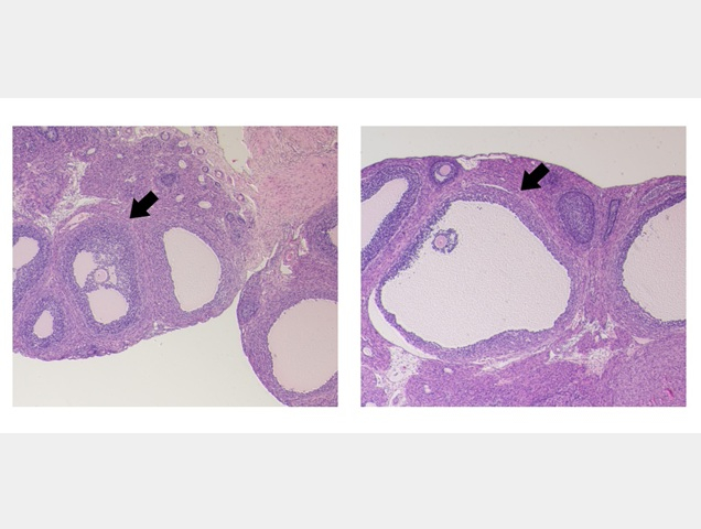
Ovaries after eCG administration
(Left) 48 hours later, (Right) 72 hours later
Credit: Osaka Metropolitan University
Researchers at Osaka Metropolitan University have found that changing the timing of fertility drugs to match the follicle maturity period significantly increases the number of ovulated oocytes—the cells that develop into eggs—during artificial fertilization, even in rats that typically produce few oocytes.
Their findings add to research on maximizing the number of oocytes. The cells are an important part of reproduction, because boosting the amount and quality that are released increases the chance of becoming pregnant.
Increasing their number in rat models usually involves hormone-based treatments, consisting of two hormones, eCG and hCG, that are administered with a 48-hour interval. In rats, eCG is often used to prime the follicles before administering hCG to induce ovulation.
However, not all rats respond the same to the treatment. Rats responsive to treatment, such as Wistar rats and F344 rats, typically release an average of 37 and 50 oocytes. On the other hand, less responsive rats like Brown-Norway (BN) rats release only seven oocytes, making them useful models for understanding why some animals do not respond to artificial fertilization.
Professor Takehito Kaneko and Dr. Yuki Nakagawa at Osaka Metropolitan University’s Graduate School of Veterinary Science successfully increased the number of oocytes retrieved following ovulation induction in BN rats by changing the timing of treatment.
The group’s discovery was based on observations of the ovaries of BN rats at 48 hours. They discovered that the follicles responsible for developing oocytes were not sufficiently mature at this time. When hCG administration was delayed to a 72-hour interval to allow the follicles to mature sufficiently, the number of oocytes ovulated following treatment increased from seven to an average of 43, similar to Wistar rats. Furthermore, 46% developed into normal offspring following fertilization, indicating normal fertilization capacity.
“Strains with low ovulation rates typically respond poorly to artificial fertilization methods, but our findings suggest that it is not that they respond poorly, but rather that follicle development is insufficient, resulting in delayed oocyte development and fewer oocytes being ovulated,” Professor Kaneko said. “We believe that the results of this study can be applied to the treatment of infertility in humans and the artificial reproduction of endangered species with low birth rates.”
The study was published in Heliyon.
Funding
This work was supported by the NIBB Collaborative Research Program, Japan [grant numbers 19-907, 20–709, 21–604, 22NIBB702, 23NIBB705, 24NIBB707 and 25NIBB702]; Environment Research and Technology Development Fund [JPMEERF20214001 and JPMEERF20244M01] of the Environmental Restoration and Conservation Agency of Japan; and Drug Discovery grant [Basis for Supporting Innovative Drug Discovery and Life Science Research, BINDS] from AMED [grant number JP22ama121049, JP23ama121049, JP24ama121049, JP25ama121049].
Paper information
Journal: Heliyon
Title: Importance of the eCG-hCG injection interval for superovulation, fertilization, and embryonic development in rats
DOI: 10.1016/j.heliyon.2025.e43619
Authors: Yuki Nakagawa, Takehito Kaneko
Published: 12 July 2025
URL: https://doi.org/10.1016/j.heliyon.2025.e43619
Contact
Takehito Kaneko
Graduate School of Veterinary Science
Email: takehito[at]omu.ac.jp
*Please change [at] to @.
SDGs
