Research News
Sep 18, 2025
- Medicine
Achalasia diagnosis simplified to AI plus X-ray
Complicated, invasive achalasia diagnosis simplified to X-rays analyzed by AI
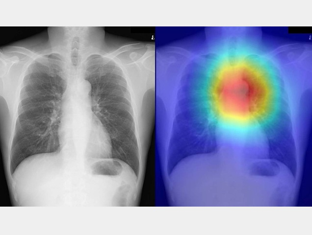
Heatmap overlay of X-ray images used for diagnosis of achalasia showing typical dilation seen in conditions like achalasia
A heatmap overlay highlighting regions of interest by the AI model. The bright red/yellow area is centered over the esophagus, indicating the model is detecting an abnormality in that region, likely dilation, a common symptom of achalasia.
Credit: Osaka Metropolitan University
Achalasia is a disease caused by impaired movement of the esophagus. Patients experience food getting stuck and regurgitated, as well as chest pain. Currently, upper gastrointestinal endoscopy and high-resolution manometry are commonly used for diagnosis; however, these techniques are invasive.
Achalasia has some distinct features that are visible on plain chest X-ray such as twisting or dilation of the esophagus, and fluid retention. However, these signs are vague in most cases, and for this reason, X-rays normally require swallowing barium to diagnose the condition.
A research group from Osaka Metropolitan University Graduate School of Medicine led by Dr. Tadashi Ochiai, Dr. Akinari Sawada, and Associate Professor Daiju Ueda created an AI model for diagnosing achalasia that uses plain chest X-ray imaging to identify the distinct features of the disease, which successfully diagnosed the disease.
The research team trained their AI model using 207 chest X-rays from 144 patients with esophageal achalasia and 240 chest X-rays from 240 age- and sex-matched non-achalasia patients. The diagnostic capability of the AI model was then verified using a test dataset consisting of 17 chest X-rays from 17 patients with esophageal achalasia and 64 chest X-rays from 64 patients without achalasia.
The diagnostic performance of the AI model was highly accurate (AUC 0.964, sensitivity 0.941, and specificity 0.891). The diagnostic performance of the AI model demonstrated higher sensitivity and specificity than physicians who reviewed the same images.
“There are reports indicating that from the onset of symptoms to diagnosis, esophageal achalasia takes an average of 6.5 years. Delayed diagnosis may worsen esophageal dilation and tortuosity and reduce treatment efficacy; therefore, early diagnosis is desirable,” Dr. Sawada said. “In Japan, chest X-rays are commonly taken during regular health checkups. Based on the findings of this study, it may be possible to screen for esophageal achalasia in a simple and minimally invasive manner.”
Paper information
Journal: Clinical Gastroenterology and Hepatology
Title: Artificial Intelligence-Based Detection of Achalasia on Plain Chest Radiography
DOI: 10.1016/j.cgh.2025.08.024
Authors: Tadashi Ochiai, Daiju Ueda, Akinari Sawada, Masaki Ominami, Akira Yamamoto, Yasuhiro Fujiwara
Published: 18 September 2025
URL: https://doi.org/10.1016/j.cgh.2025.08.024
https://www.cghjournal.org/article/S1542-3565(25)00744-X/fulltext
Contact
Akinari Sawada
Graduate School of Medicine
Email: a.sawada[at] omu.ac.jp
*Please change [at] to @.
SDGs
