Research News
Jan 11, 2024
- Medicine
New Study Unveils Machine Learning-Aided Non-Invasive Imaging for Rapid Liver Fat Visualization
The proposed framework, which is label-free and rapid, can enable an early diagnosis, treatment, and prevention of liver diseases.
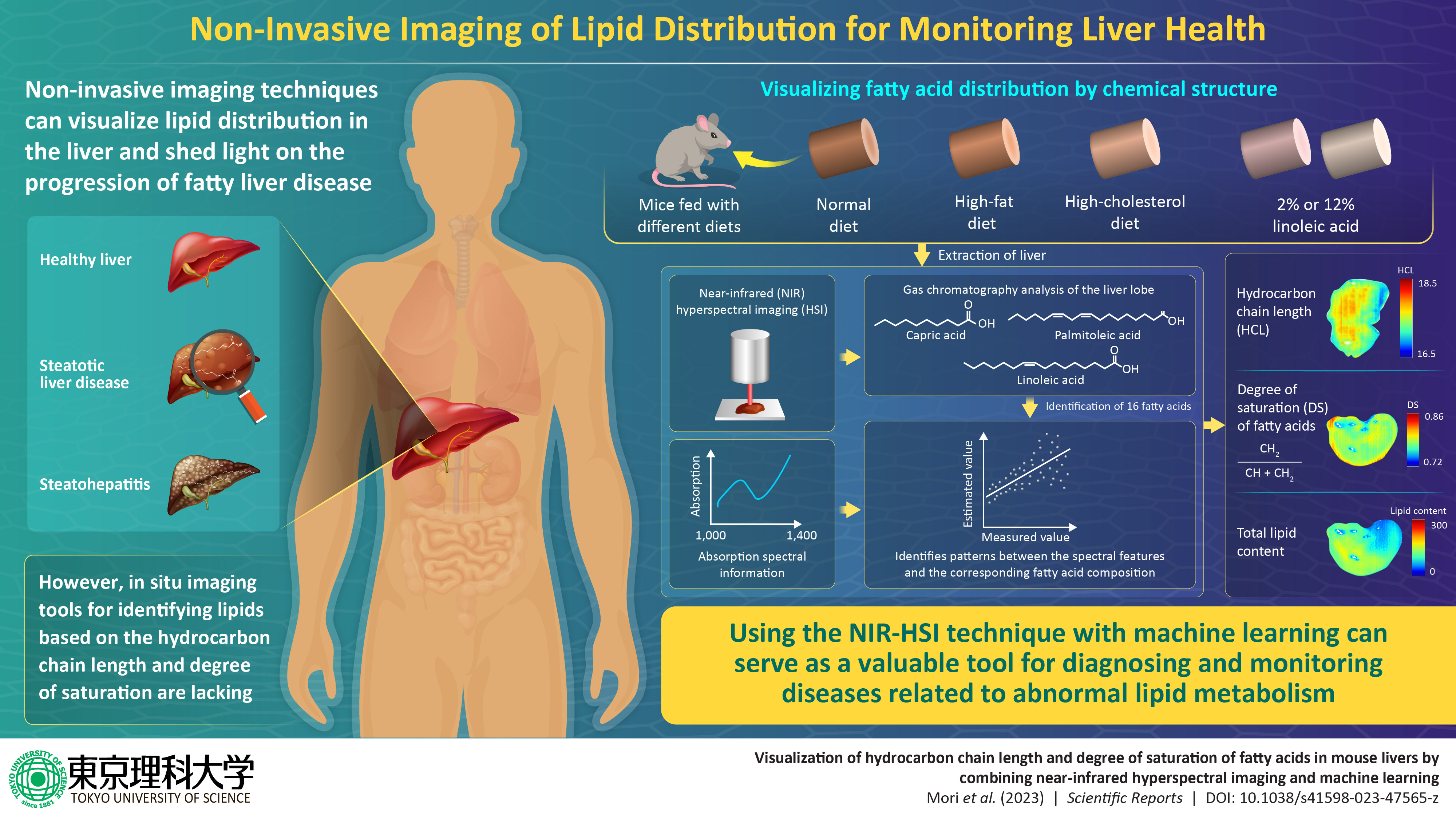
Steatotic liver disease (SLD), previously known as non-alcoholic fatty liver disease, which includes a range of conditions caused by fat build-up in the liver due to abnormal lipid metabolism, affects about 25% of the population worldwide, making it the most common liver disorder. Often referred to as “silent liver disease,” SLD progresses without noticeable symptoms and can lead to more severe conditions like cirrhosis (liver scarring) and liver cancer.
A liver biopsy—an invasive procedure involving liver tissue sample extraction from the body—is the conventional method of testing for SLD. To simplify detection, a research team led by Professor Kohei Soga of Tokyo University of Science (TUS) had previously introduced near-infrared hyperspectral imaging (NIR-HSI) as a non-invasive method to visualize the total lipid content in the liver. NIR light, with longer wavelengths (800–2500 nm) than ultraviolet and visible light shows absorption attributed to various organic substances, including biomolecules in tissues, enabling the identification of fat distribution in the liver.
Now, in a new study published in the journal Scientific Reports on 23 November 2023, the research team, including Prof. Kohei Soga, Associate Professor Masakazu Umezawa, and Associate Professor Masao Kamimura from TUS, and Professor Naoko Ohtani from Osaka Metropolitan University, has improved upon this method by having a machine learning model differentiate the type of lipids present in the liver at a pixel-by-pixel level. The framework differentiates lipids based on the hydrocarbon chain length (HCL) and degree of saturation (DS) of fatty acids, helping estimate the risk of SLD progression, steatohepatitis (NASH), and SLD/NASH-associated liver cancer.
“In addition to qualitative information, such as the total lipid content, we can now also visualize qualitative information, such as the characteristics of the distribution of fatty acids contained in lipids, mainly triglycerides,” says Dr. Umezawa.
Notably, identifying lipids based on molecular composition using NIR-HSI faced challenges due to the overlapping absorption spectra of various biomolecules. To address this, the researchers used a support vector regression machine learning model, which was trained to recognize the composition of 16 fatty acids. This training data was obtained through gas chromatography analysis of liver samples of mice that were fed diets of varying fat content. By applying machine learning to NIR-HSI data, it became possible to interpret the spectral information in terms of the distribution of fat (DS and HCL) within the liver.
DS, indicating the double bonds or degree of saturation of the fatty acids, is calculated as the CH2 fraction from the sum of the CH and CH2 numbers. HCL, representing the fatty acid chain length, is determined by the ratio of CH3 + CH2 + CH + 1(COOH) groups to the number of CH3 groups. Using this method, the researchers successfully determined the fatty acid composition in mice livers, revealing correlations with the fat contents in their diets. For instance, the livers of mice on a diet rich in saturated fats like palmitic acid and myristic acid exhibited a notably high DS, whereas mice fed with unsaturated fats such as α-linoleic acid showed a low DS.
The DS, HCL, and total lipid content were depicted as a color map, offering a unique visual representation of fat distribution in the liver, thus simplifying the diagnosis of fatty liver conditions. “Visualization of lipid distribution in higher-dimensional information rather than simply using total lipid content as a single parameter provides a novel tool for revealing the pathophysiological conditions of liver diseases and metabolism,” remarks Dr. Umezawa.
Indeed, by providing a rapid and label-free technique to identify fatty liver, which affects a large population segment, the method could be a potential alternative to invasive liver biopsy procedures, transforming liver care.
This novel framework could also find potential applications in pharmacological research, such as drug metabolism, toxicity, and efficacy; studies on metabolic disorders through metabolic imaging; and identifying responders and non-responders in clinical trials. The researchers also expect the framework to find applications in identifying personalized nutritional strategies—tailoring plans and optimizing interventions for better nutrition—through biomarker identification and treatment response prediction. In summary, the novel framework developed by the researchers could revolutionize healthcare and related research.
“Using the livers of tumor-bearing mice from our laboratory at Osaka Metropolitan University, the collaborators of Tokyo University of Science challenged a near-infrared hyperspectral imaging of the mouse livers. The research team from both universities collaborated to measure lipid contents in the mouse livers calculated by the near-infrared hyperspectral data” stated Professor Ohtani.
“This groundbreaking research used machine learning techniques to analyze the relationship between hydrocarbon chain length and saturation of lipids from actual liver lipid contents and near-infrared hyperspectral data. This research achievement enables non-invasive imaging of lipid contents, hydrocarbon chain length, and saturation status in each liver tissue. We hope this research will lead to the early detection of liver cancer and the characterization of the tissue microenvironment in the future.”

Professor Naoko Ohtani
Label-free visualization of fatty acid distribution characteristics in the liver
By combining near-infrared hyperspectral imaging (NIR-HSI) with machine learning, researchers from Japan successfully visualized the hydrocarbon chain length and degree of saturation of fatty acids in mice livers
Credit: Masakazu Umezawa from TUS, Japan
Usage restrictions: Cannot be reused without permission.
Paper Information
Journal: Scientific Reports
Title: Visualization of hydrocarbon chain length and degree of saturation of fatty acids in mouse livers by combining near‑infrared hyperspectral imaging and machine learning
DOI: 10.1038/s41598-023-47565-z
Author: Akino Mori, Masakazu Umezawa, Kyohei Okubo, Tomonori Kamiya, Masao Kamimura, Naoko Ohtani, and Kohei Soga
https://doi.org/10.1038/s41598-023-47565-z
Japanese article
https://www.omu.ac.jp/info/research_news/entry-09481.html
Contact
Department of Pathophysiology, Graduate School of Medicine
Professor: Naoko Ohtani
E-mail: naoko.ohtani[at]omu.ac.jp
*Please change [at] to @.
SDGs

